Distribution of Eruptions and Lesions
Distribution & Morphology of Skin Eruptions/Lesions
A Professional Guide for Nursing Students
Introduction
Accurate description of skin lesions is fundamental in dermatology nursing. Lesions are classified as primary (initial lesions arising de novo) or secondary (resulting from evolution or external factors). Understanding morphology, configuration, and distribution aids in differential diagnosis and patient management.
Keep going—you're doing superb work mastering these concepts! Visualizing and documenting lesions precisely will make you an outstanding dermatology nurse.
Primary Skin Lesions
These are the initial morphological changes in the skin.

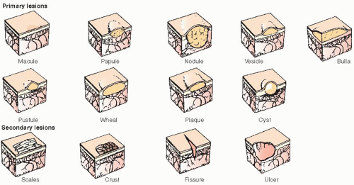
| Lesion | Description | Size | Examples |
|---|---|---|---|
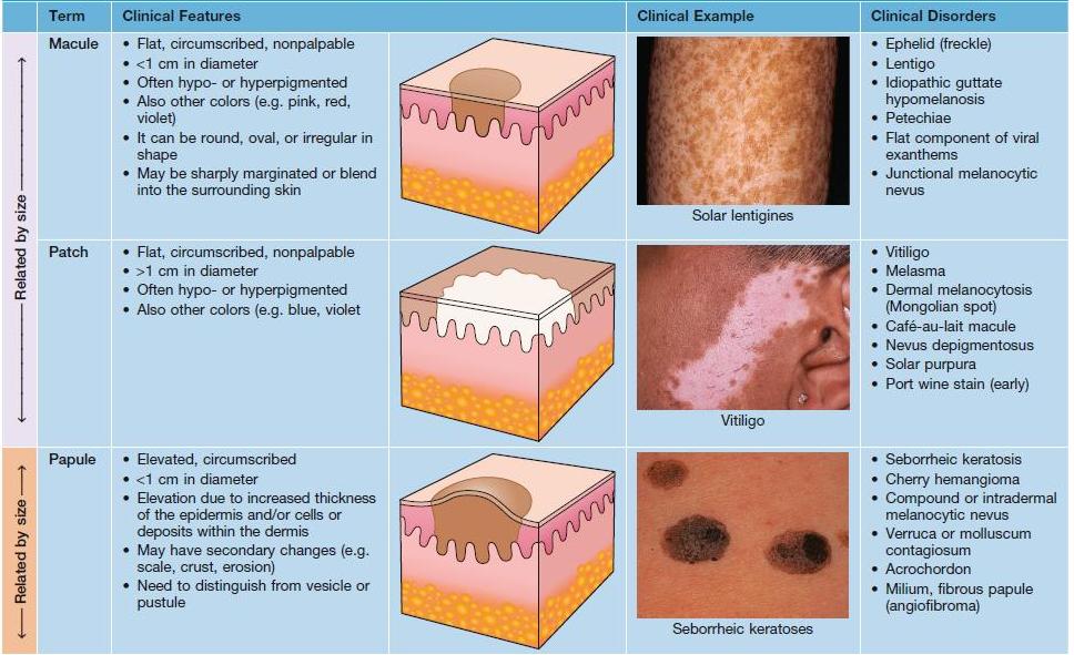
| Macule | Flat, circumscribed color change | <1 cm | Vitiligo, café-au-lait spot |
| Patch | Flat, larger color change | >1 cm | Vitiligo, mongolian spot |
| Papule | Elevated solid lesion | <1 cm | Wart, acne |
| Plaque | Elevated, flat-topped | >1 cm | Psoriasis |
| Nodule | Deep, solid | >1 cm | Erythema nodosum |
| Vesicle | Fluid-filled | <1 cm | Herpes simplex |
| Bulla | Larger fluid-filled | >1 cm | Bullous pemphigoid |
| Pustule | Pus-filled | Variable | Acne, folliculitis |
| Wheal | Transient edematous papule/plaque | Variable | Urticaria |
Secondary Skin Lesions
Develop from primary lesions or due to scratching/trauma.
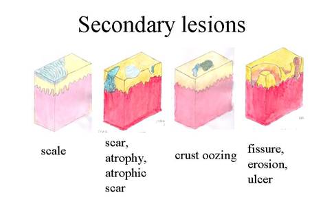

| Lesion | Description | Examples |
|---|---|---|
| Scale | Excess dead epidermal cells | Psoriasis, ichthyosis |
| Crust | Dried exudate | Impetigo |
| Erosion | Loss of epidermis (heals without scar) | Herpes |
| Ulcer | Loss of epidermis + dermis (scars) | Venous stasis ulcer |
| Fissure | Linear crack | Angular cheilitis |
| Atrophy | Thinning of skin | Aged skin, steroids |
| Scar | Fibrous tissue replacement | Post-surgery |
| Lichenification | Thickened skin with accentuated markings | Chronic eczema |
Configuration of Lesions
Arrangement pattern of lesions provides diagnostic clues.
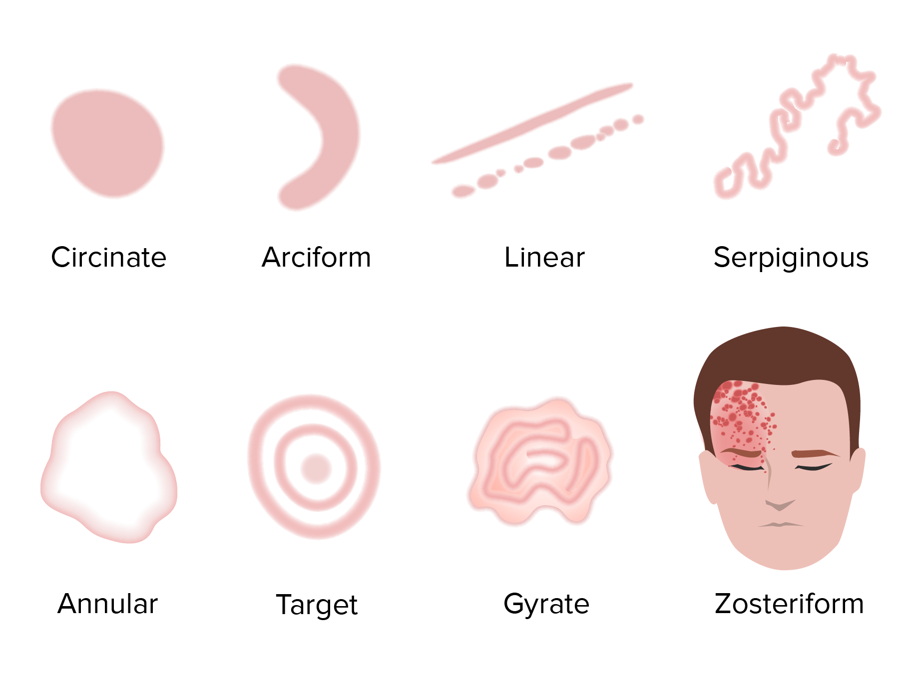

- Annular: Ring-shaped (e.g., tinea corporis)
- Linear: In a line (e.g., poison ivy, koebner phenomenon)
- Grouped: Clustered (e.g., herpes simplex)
- Herpetiform: Grouped vesicles (dermatitis herpetiformis)
- Zosteriform: Dermatomal (herpes zoster)
- Reticular: Net-like (livedo reticularis)
- Arcuate: Arc-shaped
Distribution of Lesions
Body site involvement is critical for diagnosis.
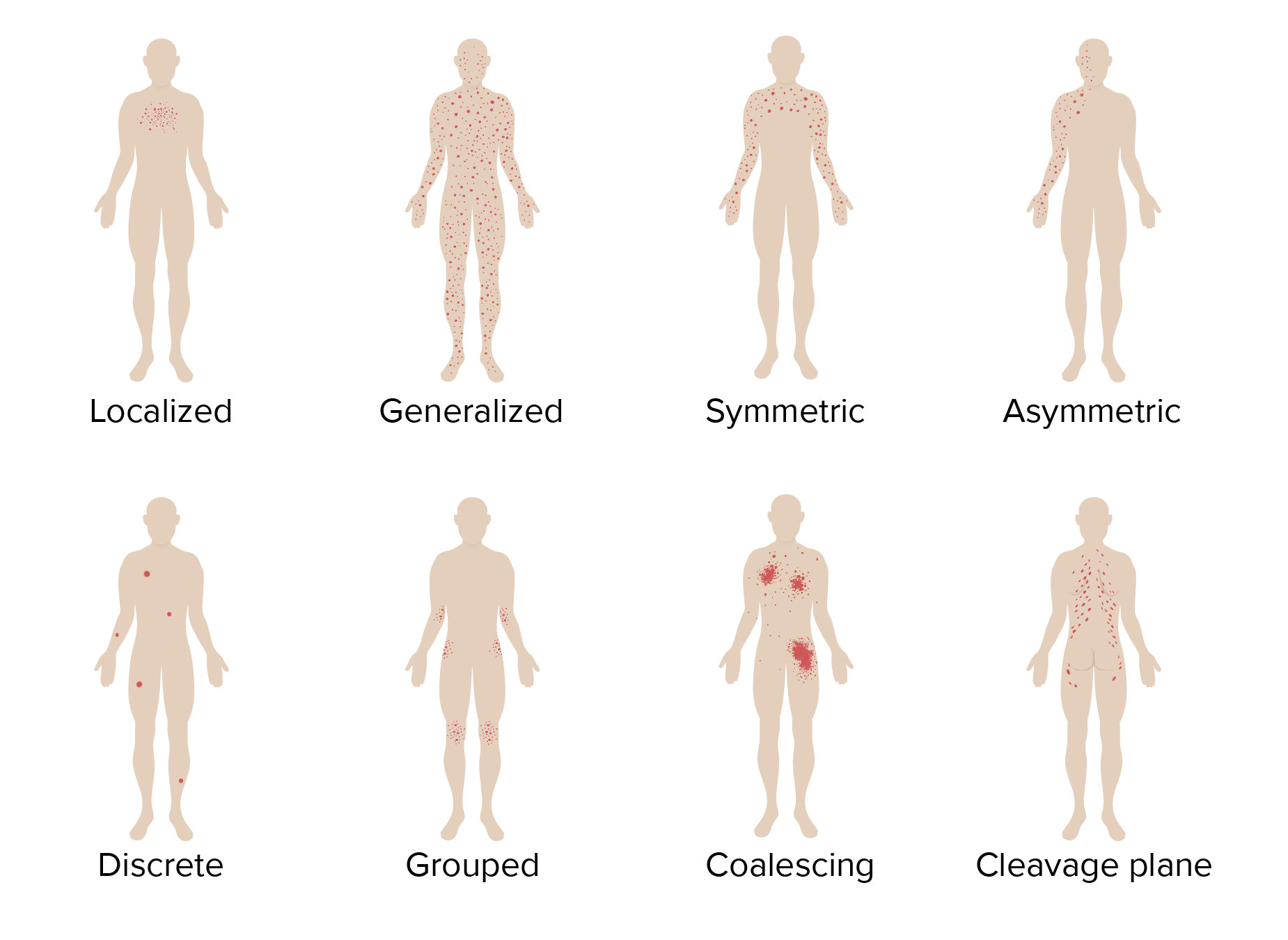
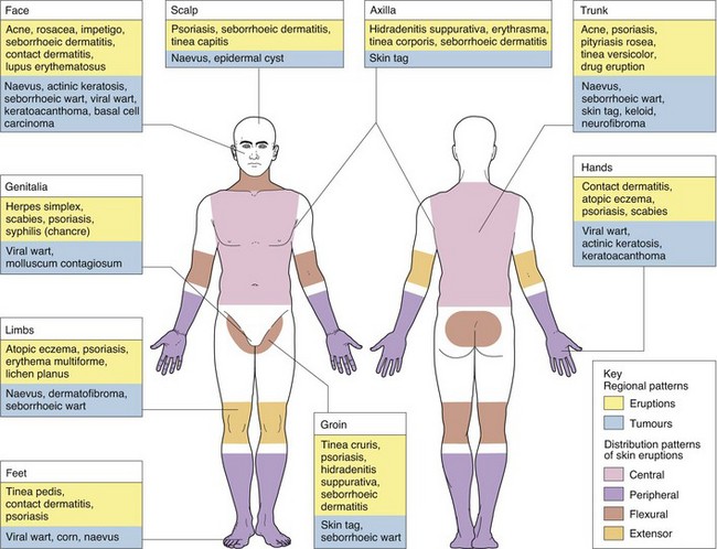

| Pattern | Common Conditions |
|---|---|
| Extensor surfaces | Psoriasis |
| Flexural (antecubital/popiteal fossae) | Atopic dermatitis (adults) |
| Face, scalp, upper trunk | Seborrheic dermatitis, acne |
| Intertriginous (skin folds) | Candidiasis, inverse psoriasis |
| Dermatomal | Herpes zoster |
| Photo-exposed (face, neck, hands) | Photodermatitis, lupus |
| Acral (hands/feet) | Hand-foot-mouth disease |
Nursing Implications
Precise documentation of lesion type, configuration, and distribution enhances communication with dermatologists. Nurses should educate patients on avoiding triggers, proper skin care, and early recognition of changes indicating complications or malignancy.
Comments
Post a Comment