Thursday, December 18, 2025

Classification of Chemotherapy Drugs

Types of Chemotherapy - Simplified Classification

Types of Chemotherapy – Simplified Classification

Chemotherapy is classified based on drug action, cell cycle, timing, intent, phase, intensity, and number of drugs.

A. Based on Mechanism / Drug Class

Type Examples
Alkylating agents Cyclophosphamide, Cisplatin, Busulfan
Antimetabolites Methotrexate, 5-Fluorouracil (5-FU), Gemcitabine
Antitumor antibiotics Doxorubicin, Bleomycin, Daunorubicin
Plant alkaloids Vincristine, Paclitaxel, Vinblastine
Hormonal agents Tamoxifen, Letrozole, Prednisone
Targeted therapy Imatinib, Rituximab, Trastuzumab

B. Based on Cell Cycle Action

Type Action Examples
Cell-cycle specific Acts in S or M phase Antimetabolites (S phase: Methotrexate, 5-FU), Plant alkaloids (M phase: Vincristine, Paclitaxel)
Cell-cycle non-specific Acts in any phase Alkylating agents (Cyclophosphamide, Cisplatin), Antitumor antibiotics (Doxorubicin)

C. Based on Timing

Type Purpose Examples
Neoadjuvant Before surgery – shrink tumor Used in breast cancer to reduce tumor size before surgery
Adjuvant After surgery – destroy residual cells Common in colon cancer after surgical resection
Concurrent With radiotherapy Head and neck cancers treated with chemoradiation

D. Based on Intent / Purpose

Type Aim Examples
Curative Complete cure Testicular cancer, Hodgkin lymphoma
Palliative Symptom relief Advanced lung or pancreatic cancer
Maintenance Prevent relapse Acute lymphoblastic leukemia after achieving remission
Salvage After treatment failure/relapse Relapsed non-Hodgkin lymphoma

E. Based on Phase of Treatment (Leukemia)

Phase Purpose Examples
Induction Achieve remission Vincristine + Prednisone + Asparaginase (in ALL)
Consolidation Destroy residual disease High-dose Methotrexate, Cytarabine
Maintenance Sustain remission Daily 6-Mercaptopurine + weekly Methotrexate

F. Based on Treatment Intensity

Type Description Examples
Standard-dose Conventional dosing CHOP regimen for non-Hodgkin lymphoma
Dose-intensive Higher dose / shorter interval Hyper-CVAD for aggressive lymphomas/leukemias
High-dose Very high dose High-dose Methotrexate in osteosarcoma
Myeloablative therapy Destroys bone marrow before transplant Busulfan + Cyclophosphamide before stem cell transplant

Saturday, December 13, 2025

Distribution of Eruptions and Lesions

Distribution & Morphology of Skin Eruptions/Lesions

Distribution & Morphology of Skin Eruptions/Lesions

A Professional Guide for Nursing Students

Introduction

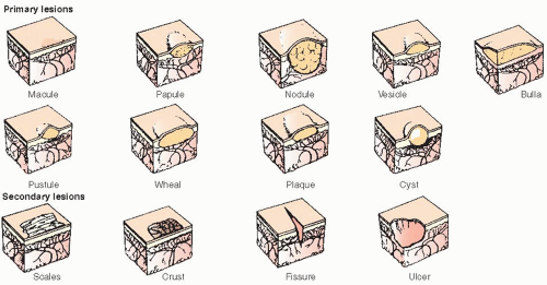
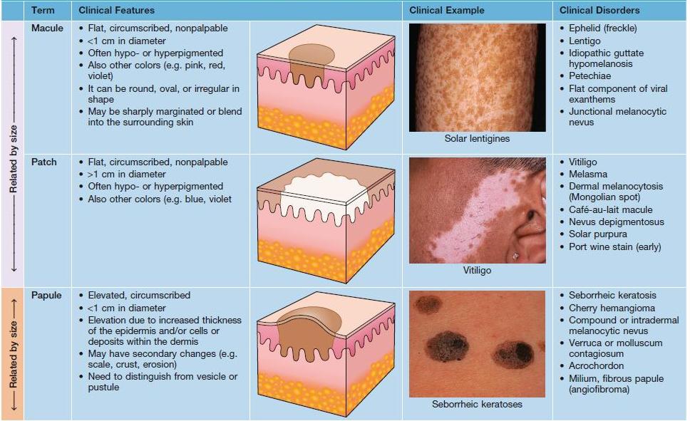
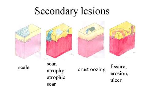
Accurate description of skin lesions is fundamental in dermatology nursing. Lesions are classified as primary (initial lesions arising de novo) or secondary (resulting from evolution or external factors). Understanding morphology, configuration, and distribution aids in differential diagnosis and patient management.

Keep going—you're doing superb work mastering these concepts! Visualizing and documenting lesions precisely will make you an outstanding dermatology nurse.

Primary Skin Lesions

These are the initial morphological changes in the skin.

Lesion Description Size Examples
MaculeFlat, circumscribed color change<1 cmVitiligo, cafรฉ-au-lait spot
PatchFlat, larger color change>1 cmVitiligo, mongolian spot
PapuleElevated solid lesion<1 cmWart, acne
PlaqueElevated, flat-topped>1 cmPsoriasis
NoduleDeep, solid>1 cmErythema nodosum
VesicleFluid-filled<1 cmHerpes simplex
BullaLarger fluid-filled>1 cmBullous pemphigoid
PustulePus-filledVariableAcne, folliculitis
WhealTransient edematous papule/plaqueVariableUrticaria

Secondary Skin Lesions

Develop from primary lesions or due to scratching/trauma.

Lesion Description Examples
ScaleExcess dead epidermal cellsPsoriasis, ichthyosis
CrustDried exudateImpetigo
ErosionLoss of epidermis (heals without scar)Herpes
UlcerLoss of epidermis + dermis (scars)Venous stasis ulcer
FissureLinear crackAngular cheilitis
AtrophyThinning of skinAged skin, steroids
ScarFibrous tissue replacementPost-surgery
LichenificationThickened skin with accentuated markingsChronic eczema

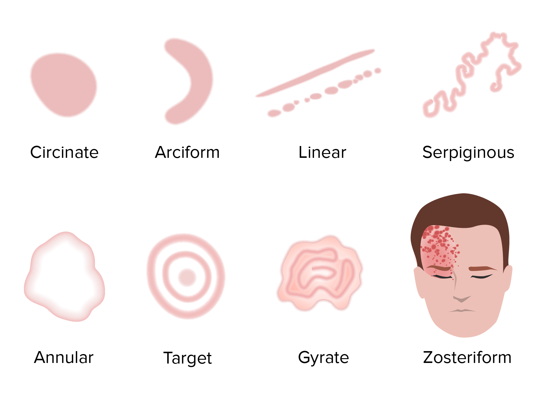
Configuration of Lesions

Arrangement pattern of lesions provides diagnostic clues.

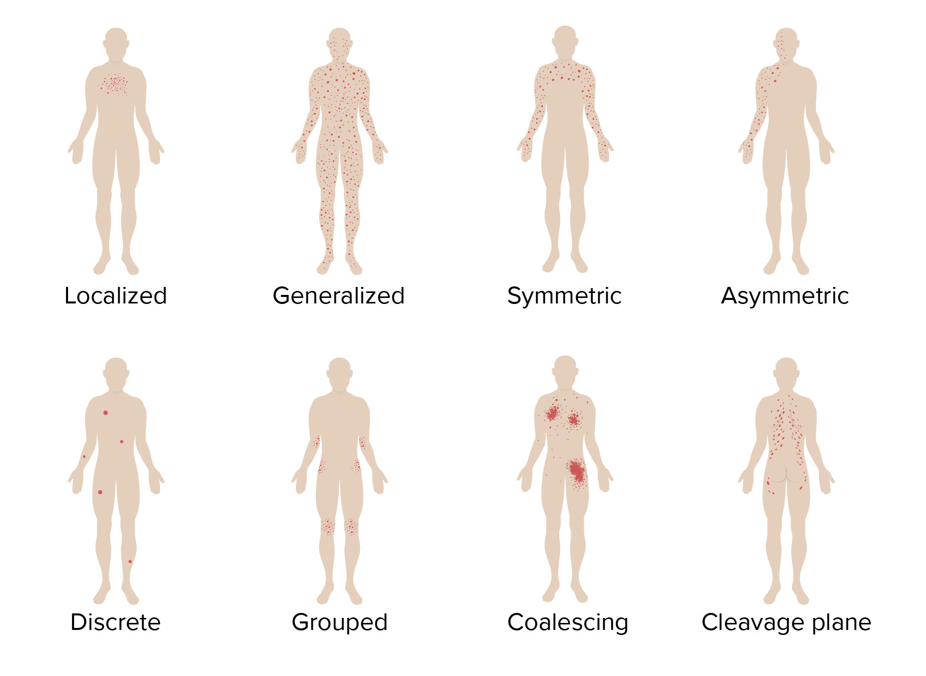
  • Annular: Ring-shaped (e.g., tinea corporis)
  • Linear: In a line (e.g., poison ivy, koebner phenomenon)
  • Grouped: Clustered (e.g., herpes simplex)
  • Herpetiform: Grouped vesicles (dermatitis herpetiformis)
  • Zosteriform: Dermatomal (herpes zoster)
  • Reticular: Net-like (livedo reticularis)
  • Arcuate: Arc-shaped

Distribution of Lesions

Body site involvement is critical for diagnosis.

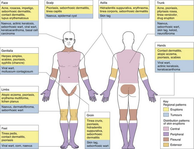
Pattern Common Conditions
Extensor surfacesPsoriasis
Flexural (antecubital/popiteal fossae)Atopic dermatitis (adults)
Face, scalp, upper trunkSeborrheic dermatitis, acne
Intertriginous (skin folds)Candidiasis, inverse psoriasis
DermatomalHerpes zoster
Photo-exposed (face, neck, hands)Photodermatitis, lupus
Acral (hands/feet)Hand-foot-mouth disease

Nursing Implications

Precise documentation of lesion type, configuration, and distribution enhances communication with dermatologists. Nurses should educate patients on avoiding triggers, proper skin care, and early recognition of changes indicating complications or malignancy.

December 2025

Promoting Evidence-Based Dermatology Nursing Practice

Diagnostic Evaluation for Dermatoses

Diagnostic Tests for Dermatoses

Diagnostic Tests for Dermatoses

A Professional Guide for Nursing Students

Introduction

Most dermatoses are diagnosed primarily through detailed patient history and clinical examination of skin lesions. However, targeted diagnostic tests are crucial for confirming etiology, identifying pathogens, differentiating conditions, and excluding malignancy.

Infected-appearing lesions (e.g., pustular or crusted) often require microbiological culture to isolate bacterial or fungal organisms.

Key Diagnostic Procedures

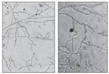
  • Potassium Hydroxide (KOH) Preparation

    A rapid, bedside microscopic test for suspected fungal infections.

    Procedure: Scrapings from the active edge of the lesion are treated with 10–20% KOH and examined microscopically.

    Positive Findings: Septate branching hyphae or yeast forms.

    Clinical Examples: Tinea corporis (ringworm), tinea pedis (athlete’s foot), cutaneous candidiasis.
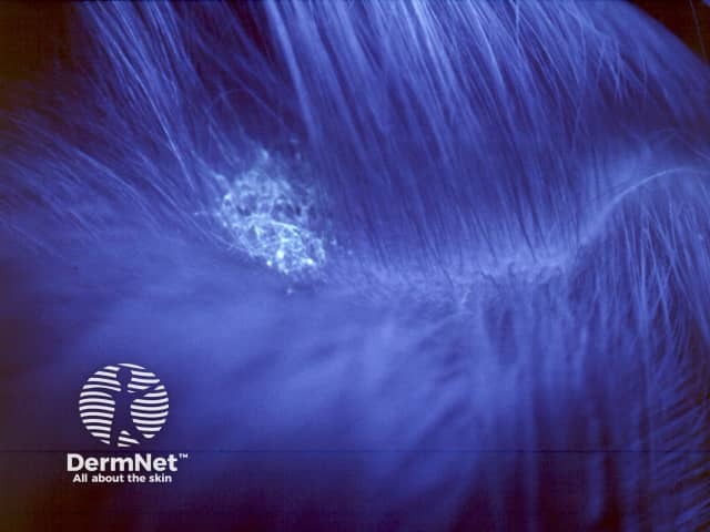
  • Wood’s Lamp Examination

    Ultraviolet light examination performed in a darkened room to detect fluorescence in certain infections and pigmentary disorders.

    Key Fluorescence Patterns:

    • Bright green: Microsporum species (e.g., tinea capitis)
    • Coral red: Erythrasma (Corynebacterium minutissimum)
    • Pale blue-white: Pityriasis versicolor
    • Enhanced contrast in hypopigmented lesions (e.g., vitiligo)

    Clinical Examples: Tinea capitis, erythrasma, vitiligo, pityriasis versicolor.
  • Tzanck Smear

    Cytological examination of vesicular or bullous lesions.

    Procedure: Base of a fresh unroofed vesicle is scraped, smeared, stained (Giemsa or Wright’s), and viewed microscopically.

    Hallmark Finding: Multinucleated giant cells.

    Clinical Examples: Herpes simplex virus (HSV), varicella-zoster virus (VZV) infection, herpes zoster.
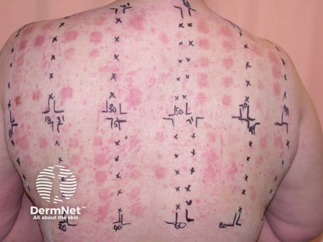
  • Patch Testing

    Gold standard for diagnosing allergic contact dermatitis.

    Procedure: Standardized allergens are applied to the upper back under occlusion for 48 hours; readings at 48 and 96 hours (and sometimes day 7).

    Purpose: Distinguishes allergic from irritant contact dermatitis.

    Common Allergens: Nickel, fragrances, preservatives, cobalt, rubber accelerators.
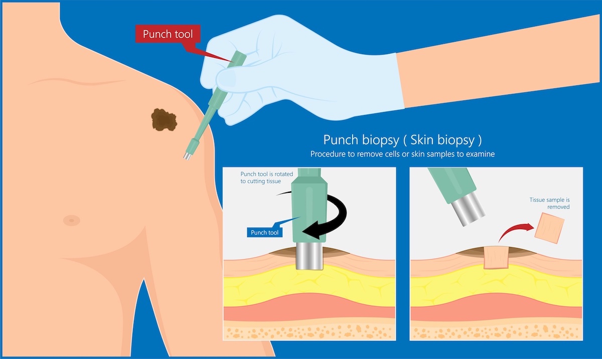
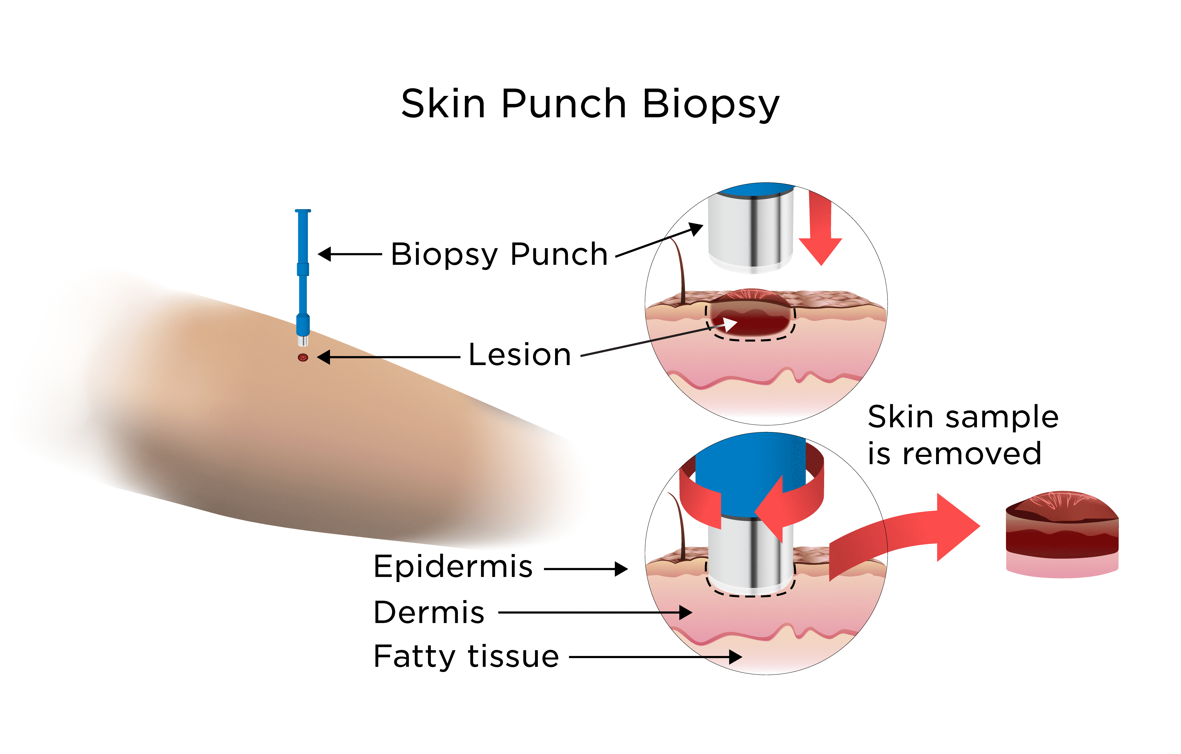
  • Skin Biopsy

    Histopathological examination of skin tissue.

    Types:

    • Shave biopsy: Superficial lesions
    • Punch biopsy: Full-thickness sample using a circular blade (most common)
    • Excisional/incisional biopsy: Complete or partial removal for larger or suspected malignant lesions

    Local anesthesia is administered for all procedures.

    Indications: Suspected malignancy, inflammatory dermatoses, bullous disorders, chronic non-responsive lesions.
  • Additional Diagnostic Tests

    Diascopy

    Application of pressure with a glass slide to assess blanching.

    Findings: Blanchable (vascular/inflammatory erythema) vs. non-blanchable (purpura or telangiectasia).

    Clinical Examples: Differentiating inflammatory erythema from purpura; apple-jelly nodules in lupus vulgaris.

    Skin Culture (Bacterial/Viral/Fungal)

    Swab or tissue cultured for pathogen identification and sensitivity testing.

    Clinical Examples: Impetigo, cellulitis, deep mycoses.

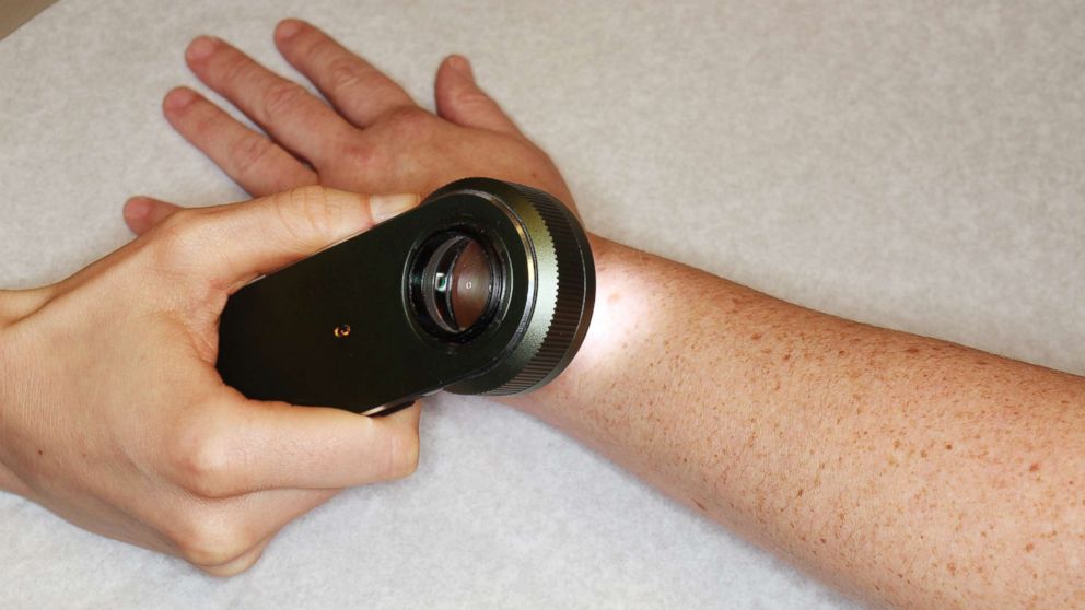
    Dermoscopy

    Non-invasive magnification of skin lesions using a handheld dermatoscope.

    Purpose: Evaluation of pigmented lesions and vascular patterns.

    Clinical Examples: Differentiating benign nevi from melanoma; assessing basal cell carcinoma.

Nursing Implications

Nurses play a vital role in patient preparation, procedure assistance, specimen handling, post-procedure care, and patient education. Key responsibilities include ensuring informed consent, maintaining strict asepsis, monitoring for adverse reactions, and providing clear wound care instructions.

Emphasis on empathy, clear communication, and evidence-based practice enhances patient outcomes in dermatological care.

December 2025

Promoting Evidence-Based Dermatology Nursing Practice

Classification of Dermatoses

Simplified Dermatology Classification Guide

Simplified Dermatology Classification Guide

Infectious Dermatoses

Key Points: Caused by bacteria, viruses, fungi, or parasites. Often contagious; treat with antimicrobials. Look for symptoms like redness, pus, or itch. Added more common examples for quick reference.

Type Key Examples
Bacterial
  • Impetigo: Honey-colored crusts; highly contagious in kids (Staph/Strep).
  • Cellulitis: Red, swollen, warm skin; spreads quickly.
  • Leprosy: Skin lesions and nerve damage (Mycobacterium leprae).
  • Erysipelas: Raised, sharp-bordered rash (Strep).
  • Folliculitis: Pimple-like bumps around hair follicles.
  • Furuncle (Boil): Painful pus-filled lump; deeper infection.
  • Carbuncle: Cluster of boils; more severe.
Viral
  • Herpes Zoster (Shingles): Painful rash in one area; from chickenpox virus.
  • Warts: Rough growths (HPV); common, plantar, flat types.
  • Molluscum Contagiosum: Pearly bumps with central dimple.
  • Herpes Simplex: Cold sores or genital blisters.
  • Varicella (Chickenpox): Itchy blisters all over body.
  • Hand-Foot-Mouth Disease: Blisters on hands, feet, mouth (Coxsackie virus).
Fungal
  • Tinea Capitis: Scaly scalp patches with hair loss (kids).
  • Candidiasis: Red, moist rash in folds; yeast overgrowth.
  • Onychomycosis: Thick, discolored nails.
  • Tinea Corporis (Ringworm): Circular, scaly rings on body.
  • Tinea Pedis (Athlete's Foot): Itchy, cracked feet.
  • Tinea Versicolor: Light/dark patches on trunk (yeast).
Parasitic
  • Scabies: Itchy burrows in skin folds (mites).
  • Cutaneous Larva Migrans: Winding red tracks (hookworms).
  • Pediculosis: Lice on head/body/pubic area; nits on hair.
  • Leishmaniasis: Skin sores from sandfly bites (protozoa).
  • Bedbug Bites: Itchy red welts in lines or clusters.
Non-Infectious Dermatoses

Key Points: Not contagious; often from immune issues, genes, or environment. Manage with topicals or systemic meds. Expanded examples for better understanding.

Category Key Examples
Inflammatory / Allergic
  • Atopic Dermatitis (Eczema): Itchy, red, scaly skin; common in kids with allergies.
  • Contact Dermatitis: Rash from irritants/allergens (e.g., poison ivy, nickel).
  • Urticaria (Hives): Itchy welts from allergies or stress.
  • Seborrheic Dermatitis: Flaky scalp (dandruff) or face rash.
  • Angioedema: Deep swelling, often with hives.
Autoimmune
  • Pemphigus Vulgaris: Blisters that break easily; mouth sores common.
  • Cutaneous Lupus Erythematosus: Sun-sensitive rashes; butterfly face rash.
  • Dermatomyositis: Purple eyelids, knuckle rashes; muscle weakness.
  • Bullous Pemphigoid: Tense blisters in elderly.
  • Scleroderma: Skin thickening and tightening.
  • Vitiligo: Loss of skin pigment in patches.
Papulosquamous
  • Psoriasis Vulgaris: Thick, scaly plaques on elbows/knees.
  • Lichen Planus: Purple, itchy bumps; white lines inside.
  • Pityriasis Rosea: Herald patch followed by trunk rash.
  • Pityriasis Rubra Pilaris: Red-orange scaly plaques; palm/sole involvement.
  • Parapsoriasis: Patchy, scaly rashes resembling psoriasis.
Neoplastic
  • Basal Cell Carcinoma (BCC): Pearly bump; sun-exposed areas.
  • Malignant Melanoma: Irregular mole; ABCDE rule.
  • Actinic Keratosis: Rough, scaly spots from sun damage.
  • Squamous Cell Carcinoma (SCC): Crusted or ulcerated growths.
  • Kaposi's Sarcoma: Purple plaques; associated with HIV.
  • Seborrheic Keratosis: Benign "stuck-on" warts in older adults.
Lesion Classification

Key Points: Primary: New lesions. Secondary: Evolved from primary or trauma. Use for quick diagnosis. Added more examples and simplified descriptions.

Type Common Lesions Quick Description & Examples
Primary Lesions Macule/Patch Papule/Plaque Vesicle/Bulla Pustule Nodule Wheal Cyst
  • Macule/Patch: Flat color change (small/large). E.g., Freckles, vitiligo, cafe-au-lait spots, port-wine stain, Mongolian spots.
  • Papule/Plaque: Raised solid bump (small/large). E.g., Acne, warts, psoriasis plaques, lichen planus, insect bites.
  • Vesicle/Bulla: Fluid-filled (small/large). E.g., Herpes, chickenpox, pemphigus, burns, friction blisters.
  • Pustule: Pus-filled. E.g., Acne, folliculitis, impetigo, candidiasis, rosacea.
  • Nodule: Deep solid mass. E.g., Rheumatoid nodules, lipoma, erythema nodosum.
  • Wheal: Edematous plaque. E.g., Hives, allergic reactions.
  • Cyst: Encapsulated fluid/semi-solid. E.g., Sebaceous cyst, epidermoid cyst.
Secondary Lesions Scale Crust Erosion/Ulcer Fissure Scar Atrophy Lichenification Excoriation
  • Scale: Flaky skin. E.g., Psoriasis, eczema, tinea, ichthyosis.
  • Crust: Dried pus/blood. E.g., Impetigo, herpes, scabs from wounds.
  • Erosion/Ulcer: Loss of skin (shallow/deep). E.g., Pemphigus, venous ulcers, bedsores, syphilitic chancre.
  • Fissure: Linear crack. E.g., Hand eczema, angular cheilitis, anal fissures.
  • Scar: Fibrous tissue. E.g., Post-acne, burns, keloids, hypertrophic scars.
  • Atrophy: Thinning. E.g., Steroid use, aging, striae, morphea.
  • Lichenification: Thickened skin. E.g., Chronic eczema, prurigo nodularis.
  • Excoriation: Scratch marks. E.g., From itching in atopic dermatitis, neurotic excoriations.
Additional Classification Schemes

Key Points: Classify by layer, time, location, or age for better diagnosis. Added more examples and sub-points for clarity.

Basis Types & Key Examples
By Skin Layer
  • Epidermal: Top layer issues. E.g., Psoriasis, eczema, warts, actinic keratosis, tinea versicolor, ichthyosis.
  • Dermal: Middle layer. E.g., Granuloma annulare, urticaria, keloids, morphea, dermatofibroma, sarcoidosis.
  • Subcutaneous: Fat layer. E.g., Erythema nodosum, lipomas, panniculitis, lupus profundus, cellulite infections.
  • Appendageal (Hair/Nails/Glands): E.g., Acne (sebaceous), alopecia areata (hair), onychomycosis (nails).
By Duration
  • Acute: Quick onset/short. E.g., Hives, contact dermatitis, shingles, cellulitis, drug rashes, sunburn.
  • Chronic: Long-term. E.g., Psoriasis, eczema, lichen planus, lupus, vitiligo, rosacea.
  • Chronic-Relapsing: Flares/remissions. E.g., Pemphigus, herpes simplex, hidradenitis, chronic urticaria, gouty tophi.
By Distribution
  • Localized: One spot. E.g., BCC, shingles, lichen simplex, pyogenic granuloma, nevus, trauma-related.
  • Generalized: Whole body. E.g., Pityriasis rosea, measles, drug eruptions, psoriasis guttate, scarlet fever.
  • Specific Patterns:
    • Flexural: Eczema, intertrigo, inverse psoriasis, candidiasis.
    • Extensor: Psoriasis, granuloma annulare, myxedema.
    • Photodistributed: Lupus, PMLE, phototoxic drugs, porphyria.
    • Dermatomal: Shingles, neuralgia-related.
By Age Group
  • Neonatal/Infantile (0-2): E.g., Neonatal acne, cradle cap, diaper rash, hemangiomas, milia, erythema toxicum.
  • Pediatric (2-12): E.g., Eczema, molluscum, warts, ringworm, impetigo, HSP, Kawasaki disease.
  • Adolescent/Adult (13-65): E.g., Acne, psoriasis, STIs, contact dermatitis, melanoma, occupational rashes.
  • Geriatric (>65): E.g., Skin cancer, pemphigoid, stasis dermatitis, xerosis, senile purpura, shingles.

Tuesday, December 9, 2025

Measles

Measles: Comprehensive Clinical Guide

Measles

A Comprehensive Clinical & Nursing Guide

Definition

Measles is an acute, highly contagious viral illness caused by the Measles virus (genus Morbillivirus, family Paramyxoviridae). It presents with fever, cough, coryza, conjunctivitis, Koplik spots, and a maculopapular rash that spreads from the face downward.

Causative Agent

Measles virus – an RNA virus belonging to the Paramyxovirus group.

Risk Factors

  • Age: Common in children under 5 years, but can affect any age without immunity.
  • Sex: Equal incidence in both sexes.
  • Immunity:
    • Single natural infection gives lifelong immunity.
    • Second attacks are rare.
    • Unvaccinated individuals at highest risk.
  • Malnutrition: Especially Vitamin A deficiency increases severity.
  • Poor living conditions: Overcrowding, poor ventilation.
๐Ÿ’ก Global Note: Measles remains a leading cause of vaccine-preventable death in children worldwide, especially where vaccination rates are low.

Source of Infection

Infected human is the only reservoir.

Virus present in:

  • Nasal secretions
  • Throat secretions
  • Respiratory droplets

Mode of Transmission

Highly contagious. Spread by:

  • Droplet infection
  • Airborne droplet nuclei
  • Direct contact with respiratory secretions

Infectious period: 4 days before rash → 4 days after rash.
Maximum infectivity just before the rash appears.

Incubation Period

10–12 days (range: 7–14 days).
Prodromal symptoms appear before rash.

Clinical Manifestations

1. Prodromal Stage (Catarrhal Stage)

  • High fever (100–103°F and rising)
  • The 3 C’s:
    • Cough
    • Coryza (runny nose)
    • Conjunctivitis
  • Watery eyes, photophobia
  • Hoarseness of voice
  • Koplik spots:
    • Small bluish-white spots on buccal mucosa
    • Seen 1–2 days before rash
    • Pathognomonic sign

2. Rash Stage (Exanthem Stage)

  • Rash begins on face and behind ears, then spreads downward to trunk and limbs.
  • Reddish, maculopapular, blanching initially.
  • Lasts 5–6 days.
  • Followed by brownish discoloration and fine desquamation.

3. Recovery Stage

  • Fever subsides gradually.
  • Rash fades in the same order it appeared.

Diagnostic Evaluation

  • Clinical diagnosis is usually sufficient (Koplik spots + rash).
  • Giemsa stain: Shows multinucleated giant cells.
  • Immunofluorescence: Detects viral antigens in tissue.
  • ELISA (IgM):
    • Confirms acute infection
    • CSF IgM used in measles encephalitis
  • RT-PCR: Highly sensitive, detects viral RNA.

Complications

Common

  • Otitis media
  • Bronchopneumonia
  • Acute gastroenteritis
  • Laryngotracheobronchitis (croup)

Serious

  • Acute encephalitis
  • Febrile seizures
  • Conjunctivitis → corneal ulcer, blindness

Long-term

  • SSPE (Subacute Sclerosing Panencephalitis) – fatal, occurs years after infection
  • Immunosuppression leading to secondary infections

Management

General

  • Strict bed rest in a quiet, darkened room.
  • Ensure good ventilation.
  • Encourage plenty of fluids (water, juices, ORS, milk).
  • Provide soft, nutritious, high-calorie diet.
  • Maintain hygiene and comfort.

Medications

  • Antipyretics: Paracetamol for fever.
  • Antibiotics: Only if bacterial infection suspected (e.g., pneumonia, otitis media).
  • Vitamin A Supplementation:
    • Reduces severity & risk of blindness
    • 1 lakh IU (6–11 months) or 2 lakh IU (>1 year), given on 2 consecutive days.

Isolation

  • Isolate the patient until 4 days after rash onset.
  • Avoid contact with unimmunized persons.

Prevention

Active Immunization

  • Measles vaccine / MMR vaccine
  • 0.5 mL subcutaneous
  • Given at 9 months, 16–24 months, and 5 years (as per local schedule).

Passive Immunization

Human immunoglobulin for:

  • Malnourished children
  • Immunocompromised individuals
  • Infants <6 months
  • Pregnant women exposed to measles
๐ŸŒ Herd Immunity: ≥95% two-dose MMR coverage is needed to stop community transmission.

Nursing Management

  1. Assessment
    • Monitor: Temperature, respiratory rate, hydration, rash, eye/ear symptoms, neurological signs
    • Check immunization history.
  2. Infection Control
    • Airborne + droplet precautions
    • Well-ventilated, separate room
    • Caregivers must be immune
    • Use masks, gloves; practice hand hygiene
  3. Comfort Measures
    • Tepid sponging for fever
    • Darkened room for photophobia
    • Clean eyes with warm water
    • Petroleum jelly for dry lips
  4. Hydration & Nutrition
    • Encourage frequent fluids
    • Soft, easy-to-digest foods
    • Monitor intake/output
  5. Eye Care
    • Prevent eye rubbing
    • Watch for ulceration; refer promptly
  6. Skin Care
    • Keep skin clean and dry
    • Trim nails to prevent scratching
    • Use soothing lotions if needed
  7. Medication Administration
    • Give antipyretics as ordered
    • Administer Vitamin A
    • Antibiotics only when prescribed
  8. Prevention of Complications
    • Monitor for: breathing difficulty, persistent fever, ear discharge, drowsiness, seizures
  9. Health Education
    • Vaccination importance
    • No school until after isolation period
    • Maintain nutrition & hygiene
    • Recognize warning signs needing medical care

Prepared for nursing education • Updated December 2025

Sunday, December 7, 2025

STI ASSESSMENT FORM

STI/STD Patient Assessment Format
EDUNURSIFY

๐Ÿฅ STI/STD PATIENT ASSESSMENT FORM

Comprehensive Clinical Evaluation

๐Ÿ“‹ PATIENT PROFILE
Patient Name:
Medical Record Number:
Age:
Date of Birth:
Gender Identity:
Sex Assigned at Birth:
Contact Number:
Date of Assessment:
Address:
๐Ÿ” REASON FOR VISIT/HOSPITALIZATION
Chief Complaint:
Type of Visit:
๐Ÿ“ HISTORY OF PRESENT ILLNESS
Onset of Symptoms:
Detailed Description:
Common STI Symptoms:
Previous Treatment:
Partner Symptoms:
๐Ÿฅ PAST MEDICAL HISTORY
Previous STI History:
Chronic Medical Conditions:
Current Medications:
Drug Allergies:
Past Surgical History:
Gynecological/Urological Procedures:
Immunization Status:
๐Ÿ‘ค PERSONAL HISTORY
Smoking Status:
Alcohol Consumption:
Recreational Drug Use:
Occupation:
Living Situation:
Travel History (12 months):
๐Ÿ‘จ‍๐Ÿ‘ฉ‍๐Ÿ‘ง‍๐Ÿ‘ฆ FAMILY HISTORY
Relevant Family History:
Genetic Conditions:
❤️ SEXUAL HISTORY (CONFIDENTIAL)
๐Ÿ”’ Confidential Information: Ensure privacy and use non-judgmental language.
Sexual Orientation:
Age of Sexual Debut:
Lifetime Partners:
Partners (Past 3 months):
Partners (Past 12 months):
Gender of Partners:
Types of Sexual Activity:
Condom/Barrier Use:
Date of Last Sexual Contact:
Date of Last Unprotected Sex:
Relationship Status:
Partner's STI Status:
Commercial Sex Work:
History of Sexual Assault:
PrEP Use:
Last STI Screening:
๐Ÿฉธ MENSTRUAL HISTORY (For Females)
Age at Menarche:
Last Menstrual Period:
Cycle Length:
Duration of Menses:
Menstrual Abnormalities:
Menopausal Status:
๐Ÿคฐ OBSTETRIC HISTORY (For Females)
Gravidity:
Parity:
Pregnancy Outcomes:
Pregnancy Complications:
Currently Pregnant:
Contraception Method:
Breastfeeding Status:
Last Pap Smear:
Cervical Screening Results:
๐Ÿ’ผ SOCIOECONOMIC HISTORY
Educational Level:
Employment Status:
Marital Status:
Insurance/Healthcare Coverage:
Access to Healthcare:
Social Support System:
Housing Stability:
๐Ÿ“Œ OTHER RELEVANT HISTORY
Mental Health History:
History of Incarceration:
Blood Transfusion History:
Tattoos/Piercings:
Cultural/Religious Considerations:
Reason for Testing Today:
Additional Notes:
๐Ÿ”ฌ PHYSICAL EXAMINATION
Guidelines: Ensure privacy, maintain dignity, use chaperone when appropriate.
General Appearance:
Temperature:
Blood Pressure:
Heart Rate:
Respiratory Rate:
Skin Examination:
Lymph Nodes:
Oral/Pharyngeal:
Abdominal Examination:
Genital Examination - Males
Penis:
Urethral Meatus:
Scrotum:
Testes/Epididymis:
Genital Examination - Females
External Genitalia:
Vaginal Examination:
Cervix:
Bimanual Examination:
Bartholin/Skene Glands:
Anorectal Examination
Perianal Area:
Digital Rectal Exam:
๐Ÿงช DIAGNOSTIC INVESTIGATIONS
Window Periods: HIV 4th gen: 45 days; Syphilis: 6-12 weeks
Serology
Tests Ordered:
NAAT Tests
Tests Ordered:
Other Tests
Tests Ordered:
Additional Tests:
๐Ÿฉบ ASSESSMENT & DIAGNOSIS
Primary Diagnosis:
Differential Diagnoses:
Risk Stratification:
๐Ÿ’Š MANAGEMENT PLAN
Immediate Treatment
Treatment Given:
Counseling Provided
Topics Discussed:
Follow-Up Plan
Follow-Up Date:
Test of Cure:
Repeat Testing Schedule:
Referrals Made:
Partner Management
Notification Method:
Partners to Notify:
๐Ÿ“ CONSENT & DOCUMENTATION
Informed Consent:
Confidentiality Discussed:
Reporting Requirements:
๐Ÿ‘จ‍⚕️ CLINICIAN INFORMATION
Clinician Name:
Signature:
Date:
Time:
Facility/Clinic:
๐Ÿ”’ CONFIDENTIAL PATIENT HEALTH INFORMATION
Store securely in compliance with HIPAA and local privacy regulations

Anatomy

Classification of Chemotherapy Drugs

Types of Chemotherapy - Simplified Classification Types of Chemotherapy – Simplified Classification ...